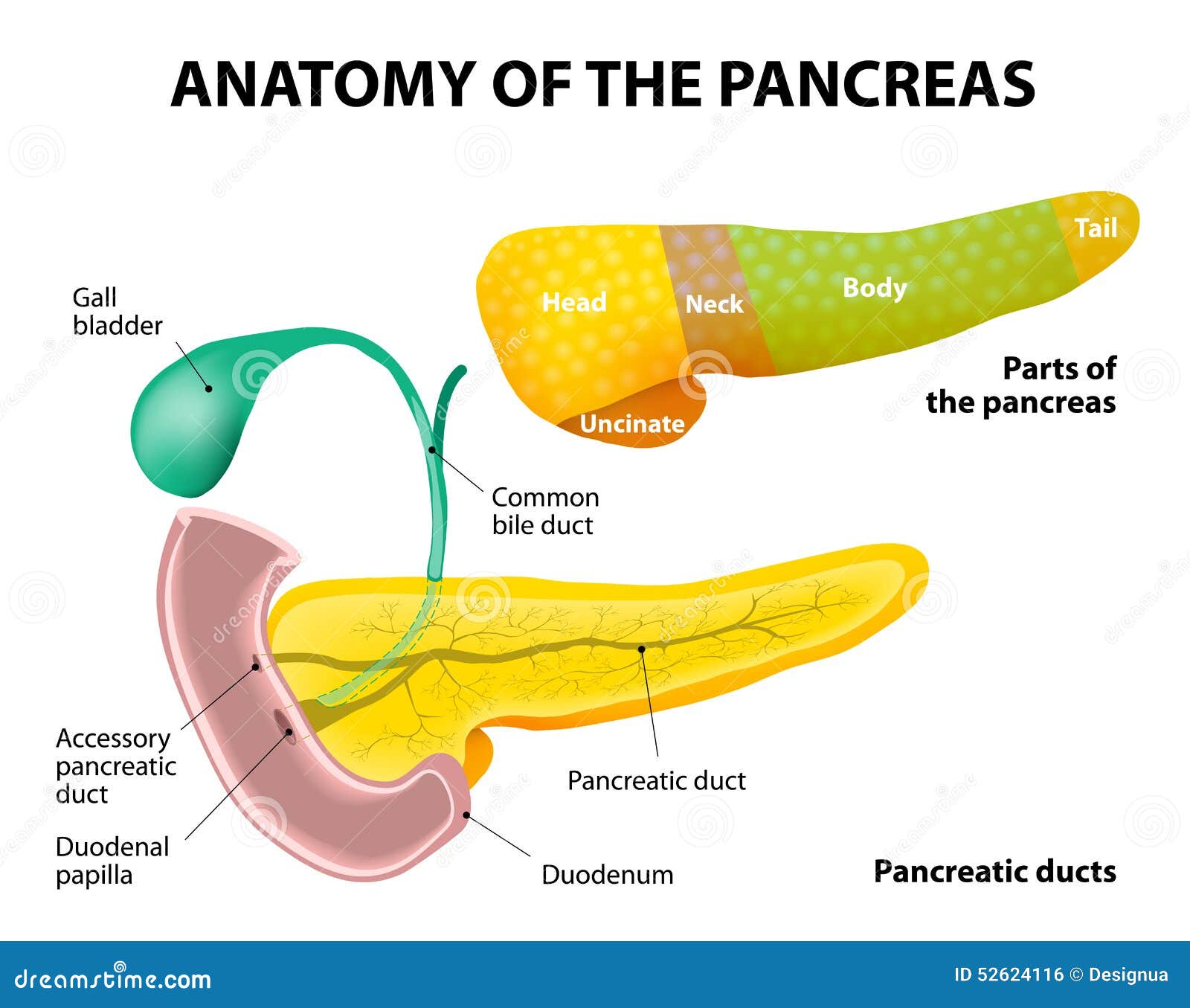
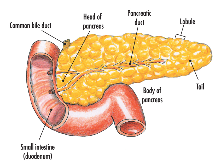
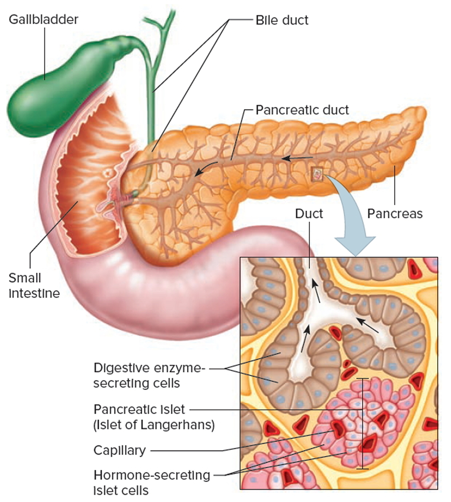
Drawing Of The Pancreas
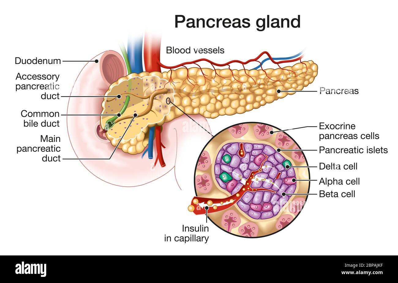
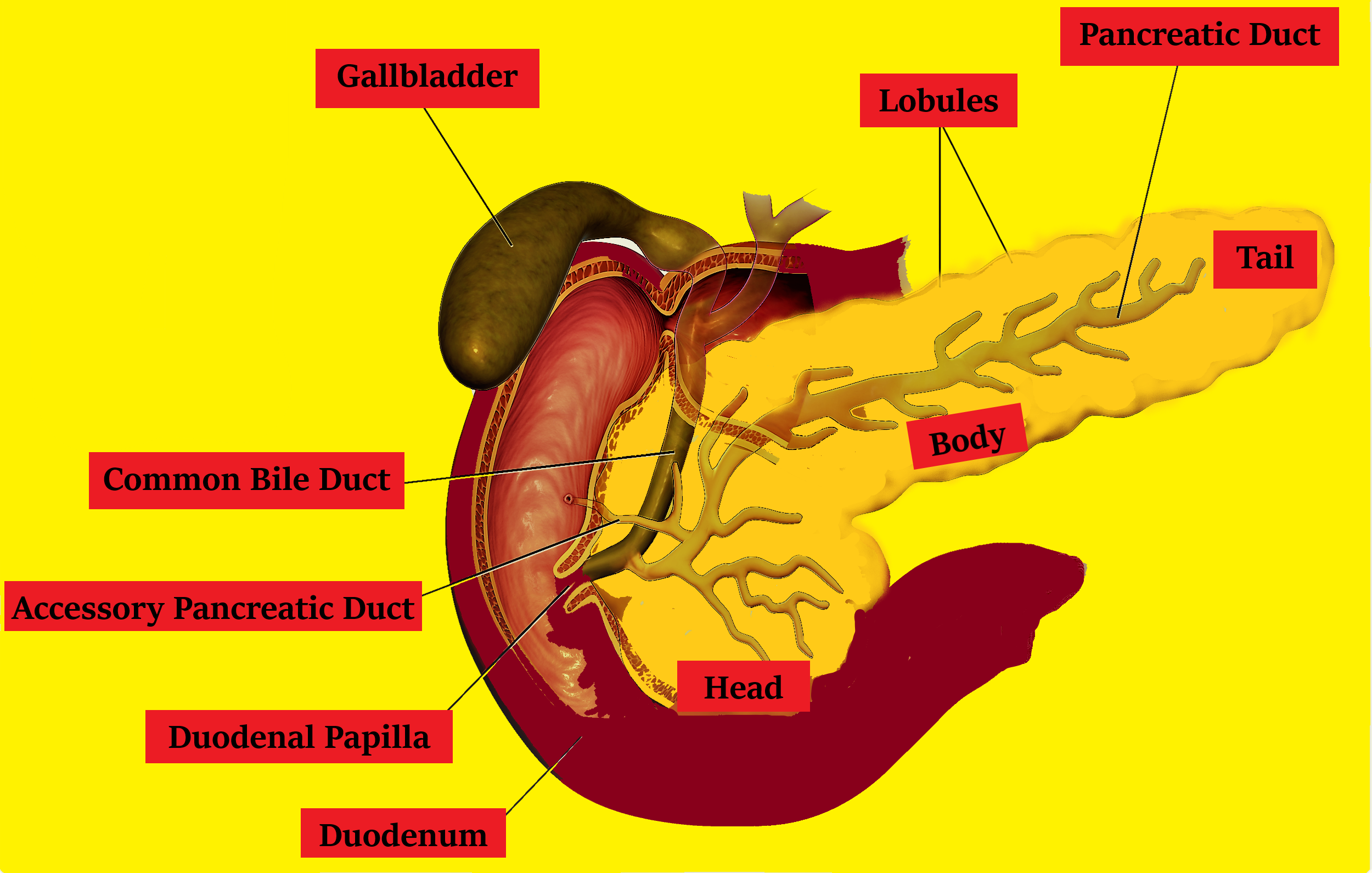
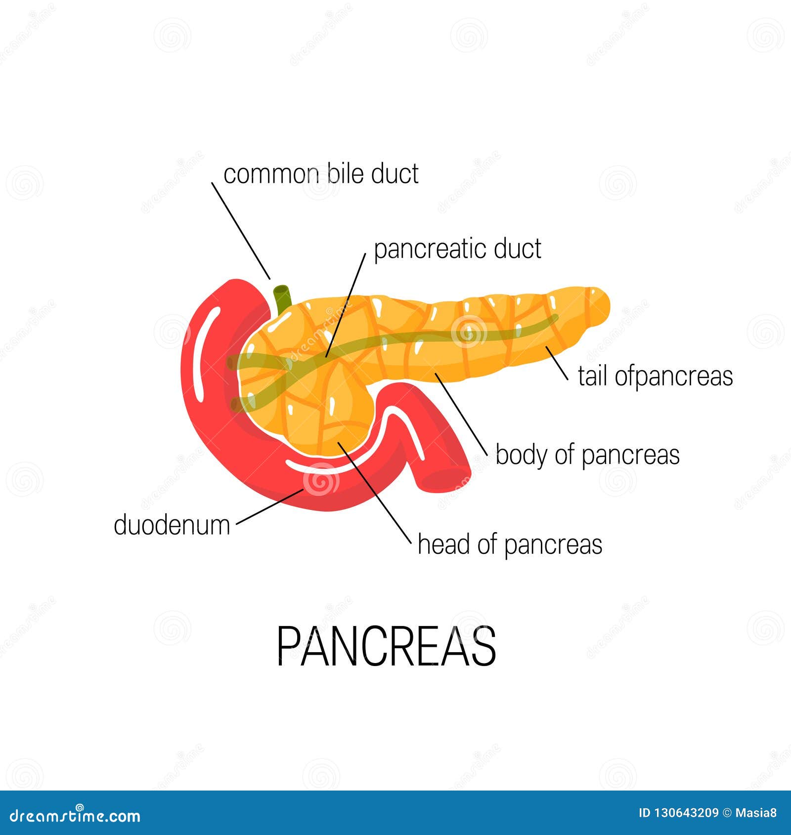
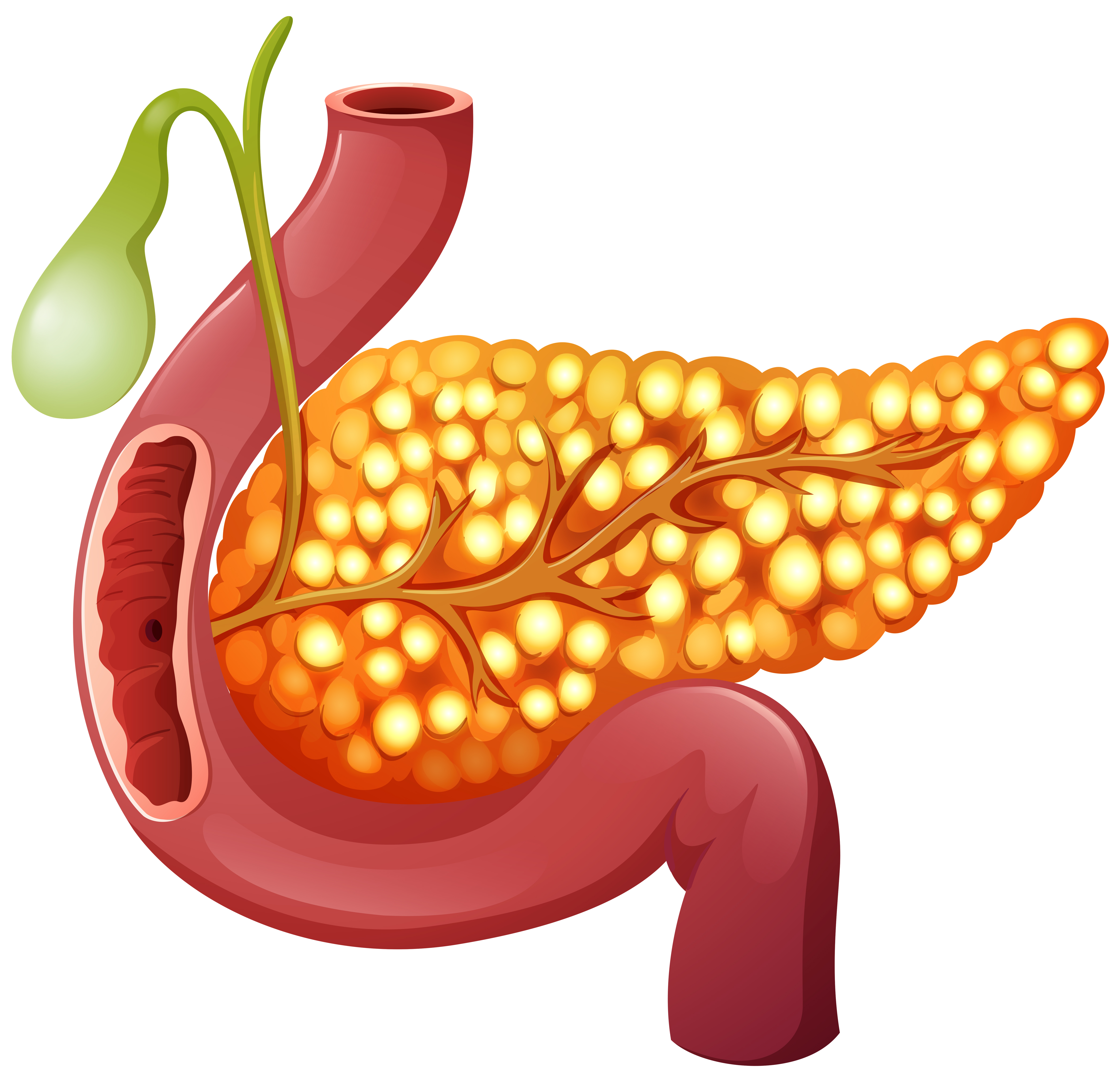
Drawing Of The Pancreas - Also, press the bell icon to get notifications. The pancreas is a gland that lies across the back side of the abdomen. Pancreatic disease can be hard to diagnose due to the location of the organ. Exocrine glandular tissues in the pancreas produce pancreatic enzymes that are dumped into the small intestine via the pancreatic duct. Please don’t forget to subscribe to my video. Web the pancreas is both an exocrine digestive gland and an endocrine gland. It serves both exocrine and endocrine functions. An inset shows the head, body, and tail of the pancreas. Human organs and anatomy related objects and elements. Compare and contrast the function and regulation of. Web these islet cells produce and secrete hormones that regulate glucose, lipid and protein metabolism. The pancreas is part of the gastrointestinal system that makes and secretes digestive enzymes into the intestine, and also an endocrine organ that makes and secretes hormones into the blood to control energy metabolism and storage throughout the body. “what we do allows us to. Please don’t forget to subscribe to my video. Web the pancreas is part of the gastrointestinal system that makes and secretes digestive enzymes into the intestine, and also an endocrine organ that makes and secretes hormones into the blood to control energy metabolism and storage throughout the body. Without it, your body can’t properly operate many. Compare and contrast the function and regulation of. The bile duct and pancreatic duct are also shown. To put it in a clinical context, its oblique position makes it impossible to see the entire pancreas in a single transverse section. Web the pancreas is viewed from the front and a portion of the parenchyma has been dissected away to reveal (1) the main (principal) pancreatic duct (wirsung’s duct) with multiple branches, (2) the accessory duct (santorini’s duct), and (3) the distal common bile duct. The pancreas extends laterally and superiorly across the abdomen from the curve of the duodenum to the spleen. Please don’t forget to subscribe to my video. Web the pancreas is an elongated gland located deep within the abdomen, tucked in between the stomach and the spine. Web anatomy of the pancreas; It secretes digestive enzymes into ducts that lead to. One end of the pancreas is wider than the other and is called the head: Web the pancreas (meaning all flesh) lies in the upper abdomen behind the stomach. Also, press the bell icon to get notifications. No matter the case or the court, the focus of sanford law's criminal defense clinic is the client as well as the alleged. The pancreas is part of the gastrointestinal system that makes and secretes digestive enzymes into the intestine, and also an endocrine organ that makes and secretes hormones into the blood to control energy metabolism and storage throughout the body. This article will describe the histology and functions of the pancreas, including a clinically relevant condition that you have definitely heard. Describe the location and structure of the pancreas, and the morphology and function of the pancreatic islets; Exocrine glandular tissues in the pancreas produce pancreatic enzymes that are dumped into the small intestine via the pancreatic duct. This article will describe the histology and functions of the pancreas, including a clinically relevant condition that you have definitely heard about, called. Human organs hand drawn line icon set. Web the pancreas is viewed from the front and a portion of the parenchyma has been dissected away to reveal (1) the main (principal) pancreatic duct (wirsung’s duct) with multiple branches, (2) the accessory duct (santorini’s duct), and (3) the distal common bile duct. Exocrine glandular tissues in the pancreas produce pancreatic enzymes. Web the pancreas is an abdominal glandular organ with both digestive (exocrine) and hormonal (endocrine) functions. It sits within the curve of the duodenum (the first part of the small intestine) and is divided into two parts: Web getting to the heart of criminal defense. An inset shows the head, body, and tail of the pancreas and the bile duct. The bile duct and pancreatic duct are also shown. It plays an important role in digestion and blood sugar regulation. Web the pancreas (meaning all flesh) lies in the upper abdomen behind the stomach. The pancreas secretes fluids that help break down food in the small. Drawing shows the pancreas, stomach, spleen, liver, bile ducts, gallbladder, small intestine, and colon. Human organs and anatomy related objects and elements. Human organs hand drawn line icon set. But there are things you can do to reduce your risk for these conditions. It controls important hormone and enzyme secretion. Drawing shows the pancreas, stomach, spleen, liver, bile ducts, gallbladder, small intestine, and colon. It sits within the curve of the duodenum (the first part of the small intestine) and is divided into two parts: Web the pancreas is part of the gastrointestinal system that makes and secretes digestive enzymes into the intestine, and also an endocrine organ that makes and secretes hormones into the blood to control energy metabolism and storage throughout the. Head, neck, body, and tail. An inset shows the head, body, and tail of the pancreas. Web the pancreas is both an exocrine digestive gland and an endocrine gland. It sits within the curve of the duodenum (the first part of the small intestine) and is divided into two parts: The pancreas extends laterally and superiorly across the abdomen from. Web the pancreas is an elongated organ (approximately 15 cm) which lies obliquely across the posterior abdominal wall, at the level of the l1 and l2 vertebral bodies. Drawing shows the pancreas, stomach, spleen, liver, bile ducts, gallbladder, small intestine, and colon. Drawing shows the pancreas, stomach, spleen, liver, bile ducts, gallbladder, small intestine, and colon. The pancreas extends laterally and superiorly across the abdomen from the curve of the duodenum to the spleen. Pancreatic disease can be hard to diagnose due to the location of the organ. Web the pancreas is both an exocrine digestive gland and an endocrine gland. It plays an important role in digestion and blood sugar regulation. Web explain the role of the pancreatic endocrine cells in the regulation of blood glucose. Human organs and anatomy related objects and elements. The pancreas is a gland that lies across the back side of the abdomen. Compare and contrast the function and regulation of. The bile duct and pancreatic duct are also shown. The pancreas is divided into 4 parts: An inset shows the head, body, and tail of the pancreas. Web the gross, histologic, and ultrastructural anatomy of the pancreas is summarized and illustrated with 33 images that include drawings, photographs, ct scans, and light and electron micrographs. Please don’t forget to subscribe to my video./pancreas_lg-595e8eae3df78c4eb64f2fce.jpg)
Pancreas Anatomy and Function
Pancreas Anatomy. labeled stock vector. Illustration of digestion
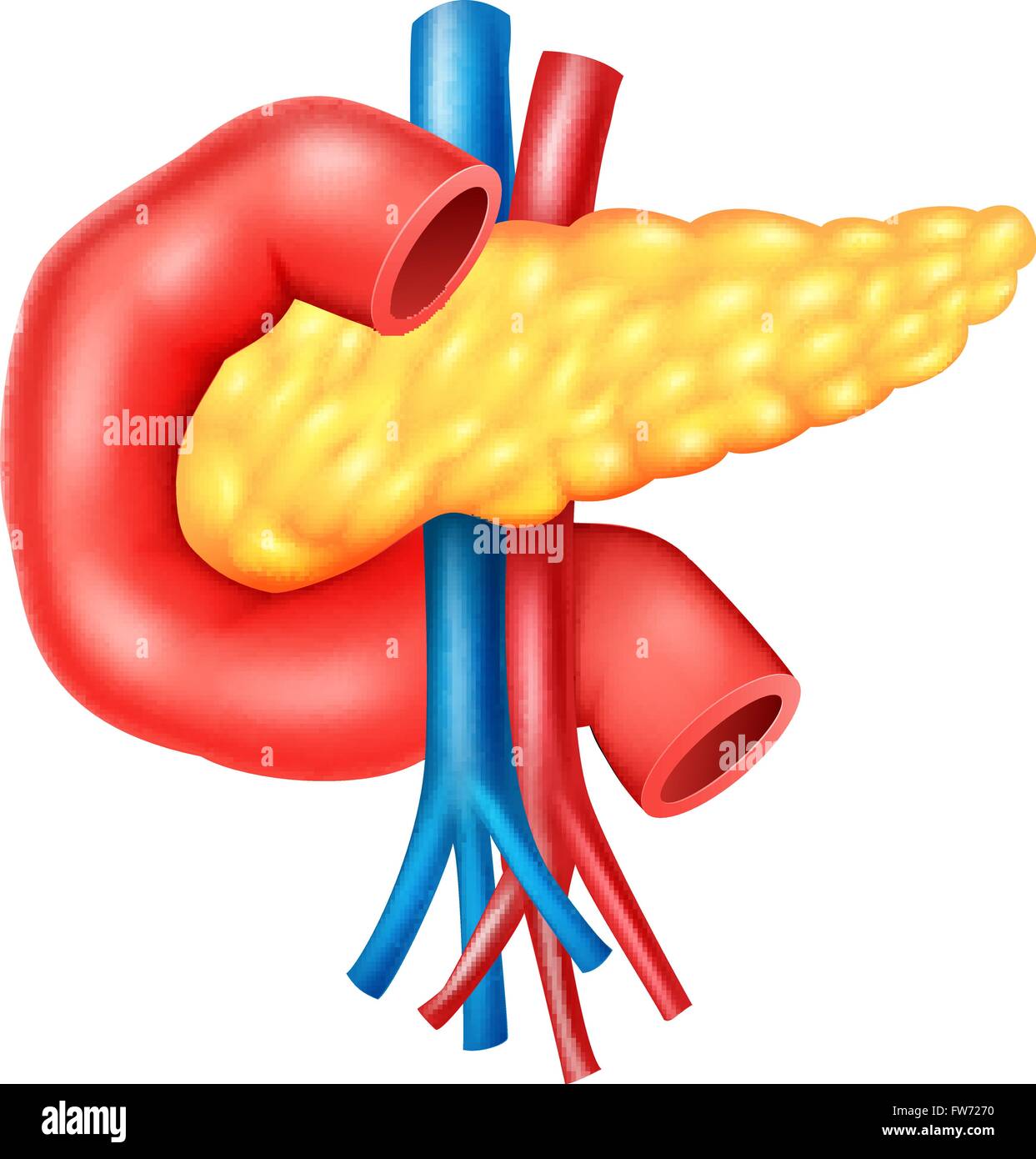
Illustration of Human Internal Pancreas Anatomy Stock Vector Image
The pancreas Anatomy of the pancreas Structure of the pancreas
Pancreas Location, Anatomy and Function in Digestion
Medically illustration showing pancreas gland and pancreatic islets
Draw a neat labeled diagram of the pancreas with their associated
Medical Diagram of Pancreas, Vector Illustration Stock Vector
A healthy human Pancreas 303570 Vector Art at Vecteezy

Human Pancreas drawing How to draw human Pancreas Human pancreas
It Serves Both Exocrine And Endocrine Functions.
An Inset Shows The Head, Body, And Tail Of The Pancreas And The Bile Duct And Pancreatic Duct.
“What We Do Allows Us To.
To Put It In A Clinical Context, Its Oblique Position Makes It Impossible To See The Entire Pancreas In A Single Transverse Section.
Related Post: